The Importance of Cross Sections in Material Analysis with SEM

Understanding the internal structure of materials is crucial for advancing product development, ensuring quality control, and solving complex engineering challenges. Cross sections provide a window into the internal layers, interfaces, and microstructural details of materials, offering critical insights into their properties and performance.
For instance, layered structures such as coated materials, multilayer composites, or lithium-ion battery electrodes often require detailed examination of layer thickness, adhesion, porosity, or potential delamination. Cross sections reveal these features, enabling researchers to understand how manufacturing processes or environmental conditions impact the material.
However, preparing high-quality cross sections for scanning electron microscopy (SEM) analysis is not without its challenges. Conventional methods like mechanical polishing often fail to produce artifacts-free surfaces, especially for delicate, heterogeneous, or highly sensitive materials. This is where advanced techniques come into play:
Cutting with Scissors or Razor Blade
Cutting with scissors or a razor blade is a straightforward and quick method, often used for initial investigations. However, this approach can introduce artifacts such as uneven edges, compression of softer materials, or delamination of layers. These artifacts may obscure finer structural details, making this method less suitable for high-precision analysis.

Embedding and Polishing
Embedding the sample in resin followed by mechanical polishing is a common technique for preparing cross sections. While effective for many materials, this method can lead to smearing of softer components, pull-out of particles, or rounding of edges. Careful optimization of polishing parameters and materials can mitigate these issues, but the risk of artifacts remains for complex or heterogeneous samples.

Scratches and marks after polishing a stainless steel using 1 um diamond
Freeze Fracture
Freeze fracture involves freezing a sample, often in liquid nitrogen, and then fracturing it to reveal internal structures. This method is particularly useful for brittle materials, biological samples, or materials with complex internal structures. While freeze fracture can preserve fine details, it may introduce artifacts such as surface roughness, uneven fracture planes, or loss of softer components during fracturing. Additionally, the preparation process can be challenging for materials that are sensitive to rapid cooling or prone to thermal stress.

Fiber enforced polymer after freeze fracture
Broad Beam Argon Milling
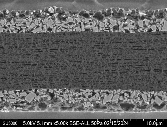
Broad beam argon milling is a versatile method that removes material gently and uniformly. It is particularly suitable for large-area cross sections of metals, ceramics, and polymers. By avoiding the mechanical stresses associated with polishing, argon milling minimizes surface damage and introduces fewer artifacts. For example, this method is excellent for analyzing the layered structure of coatings or the interfaces in composite materials.

Lithium Ion Battery separation foil cross sectioned at -90C in BIB
Focused Ion Beam (FIB)
Focused ion beam (FIB) systems excel in high-precision cross sectioning and site-specific sample preparation. They are widely used for nanostructured materials or when specific regions of interest, such as cracks or defects, must be targeted. FIB is very site specific and can be used to prepare specific points of interest with almost nm precision but on the other hand need long preparation time for larger areas.

Site specific cross section of a LiB cathode using FIB
Short-Pulsed (Femtosecond) Laser Processing
Short-pulsed femtosecond lasers offer rapid and precise cross sectioning of hard or brittle materials. Their ultra-short pulse duration minimizes thermal damage, making them ideal for processing complex multilayer structures or high-value samples where pristine preservation of the microstructure is critical. For instance, researchers studying photovoltaic cells or semiconductor devices benefit significantly from this technique.

Fs-laser cut cross section of a LiB Cathode
Integrated Microtome in SEM Vacuum Chamber
For soft or biological materials, an integrated microtome within the SEM vacuum chamber provides an innovative solution. This approach enables ultra-thin slicing and immediate SEM imaging without exposing the sample to atmospheric conditions, ensuring minimal contamination or distortion. It is a powerful tool for investigating polymer laminates or biomaterials with layered architectures. Sequential cutting and imaging allows for 3D reconstruction of mm sized volumes.

3D reconstruction after Serial Block Face (SBF) imaging using an integrated microtome in the SEM.
Conclusion
Selecting the appropriate cross sectioning technique depends on the material’s properties, the scale of features to be analyzed, and the study’s objectives. Whether uncovering the secrets of lithium-ion battery electrodes or evaluating the integrity of coatings, high-quality cross sections are fundamental to revealing the stories materials have to tell. Modern methods like cutting, embedding/polishing, freeze fracture, argon milling, FIB, femtosecond lasers, and integrated microtomes empower researchers to push the boundaries of SEM analysis, delivering accurate and reliable results.
By leveraging these advanced techniques, you can enhance your material analysis workflows and gain a deeper understanding of your samples, ultimately driving innovation and performance in your field.

